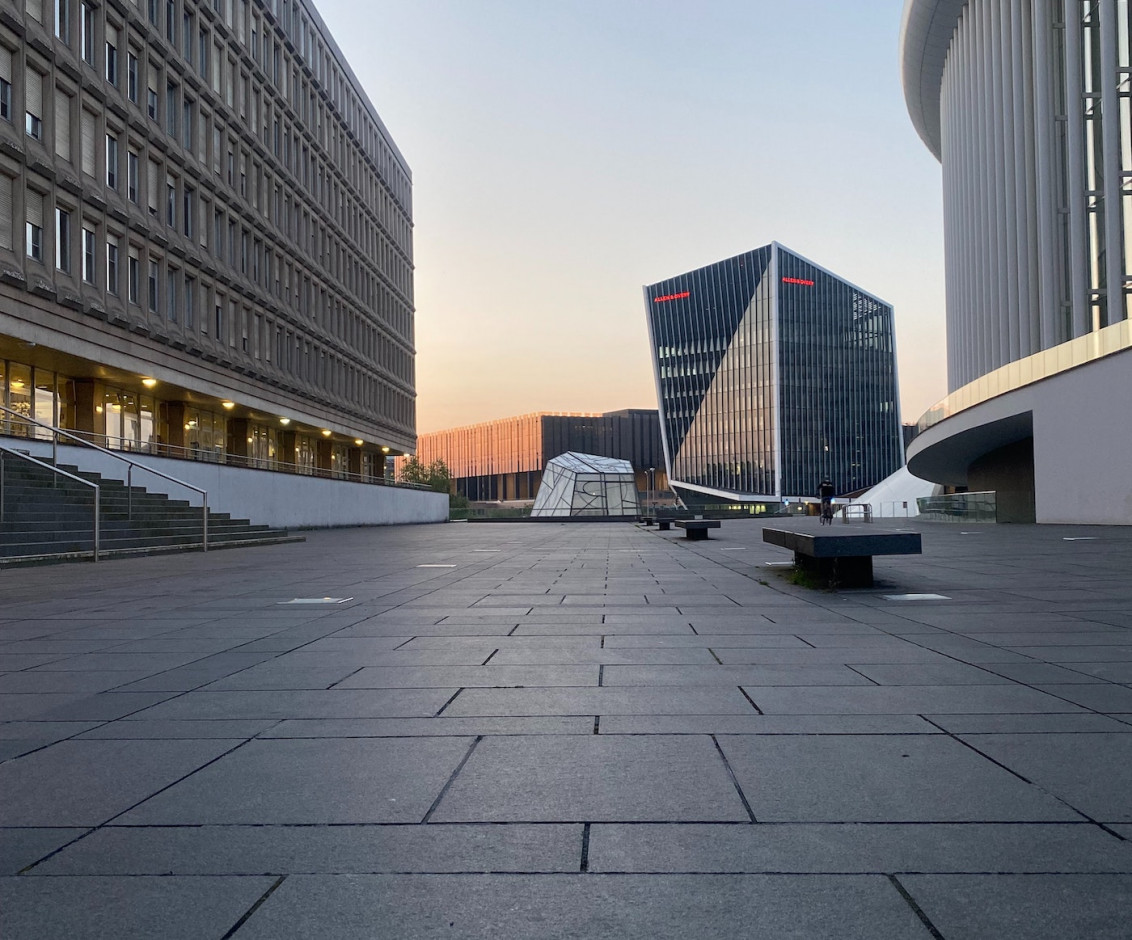
Your practice for vascular surgery in the heart of Luxemburg

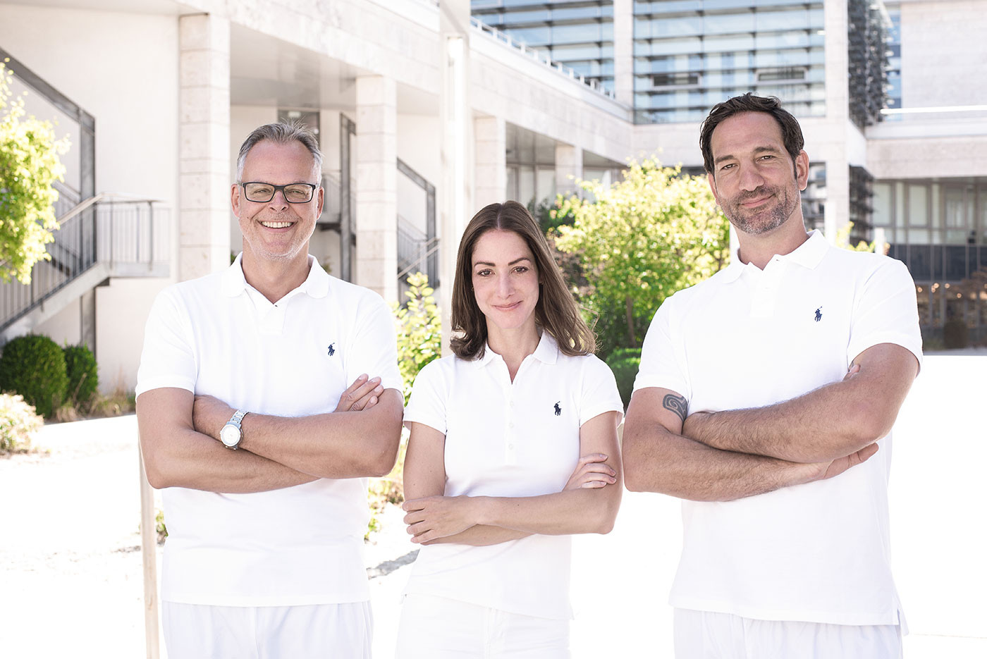
You and your health are the focus of our attention - your doctors at Chirurgie Vasculaire Kirchberg. As specialists in vascular surgery who have been practising for many years, we are characterised by an exceptionally high level of experience, competence, quality and personality.

In the Department of Vascular Surgery at the Hôpital Kirchberg, we offer our patients the latest diagnostic and therapeutic procedures as well as a comprehensive range of services at the highest medical level.

We would like you to feel in good hands and always strive for the best individual solution for the benefit of your health. To this purpose, we take a lot of time and care for you continuously from the first examination to the successful treatment.

Contact

Chirurgie Vasculaire Kirchberg
Hôpital Kirchberg
9, rue Edward Steichen
L-2540 Luxemburg

Phone +352 24684160
(08h00-17h00)
Fax +352 24684163

info@chirurgievasculaire.lu

Consultation hours

Mon 13h00-17h00 (Dr. Grotemeyer)

Tue 10h30-13h00 (Dr Manzoni)

Wed 7h00-10h30 (Dr. Kröger)

Wed 13h00-15h00 (Dr. Grotemeyer)

Wed 15h00-17h00 (Dr. Kröger)

Thu 8h00-12h30 (Dr. Kröger)

Thu 12h30-17h00 (Dr Manzoni)

And by arrangement

How to get to us

Access by car:

  • From the east and south A1, from the north A7, then exit 8 in the direction of Luxemburg-Centre, turn right in the direction of Bd Pierre Werner, follow until Rue Joseph Leydenbach, there is the entrance to the underground car park of the "Hôpital Kirchberg"

  • From the West/Centre: N1 or N51 to Rue Joseph Leydenbach, there is the entrance to the underground car park of the "Hôpital Kirchberg"

Access by public transport:

  • Bus: line 7, 12, 21, 26, 248, bus stop "Hôpital Kirchberg", 88 metres away, 2 min walking distance

  • Bus: Line 11 and 303 to stop "Kirchberg, Hugo Gernsback", 650 metres away, 9 min walking distance

  • Tram: Stop "Alphonse Weicker", 477 metres away, 7 min walking distance

  • Tram: The Glacis Luxemburg car park (1€ per hour) is 270m from the tram stop "Limpertsberg, Theater", from there 7 stops (11min) to the stop "Alphonse Weicker"

Make an appointment

Doctor
Are you already our patient?

Thank you for your enquiry.

We will get back to you shortly.

Specialists in vascular medicine who love their job

Our Doctors

Priv.-Doz. Dr. med. Dirk Grotemeyer

Training

  • Paramedic training

    Dortmund, Germany 02/1986-09/1987

  • Study of human medicine

    Medical University Duesseldorf, Kiel und Aachen, Germany 10/1987-10/1994

  • Study Health Economics

    European Business School ebs, Schloss Reichartshausen, Oestrich-Winkel at Rheingau, Germany 06/1999-04/2000

Medical practice

  • Service de Chirurgie Vasculaire

    Hôpital Kirchberg, Luxemburg, since 01/2010

  • Senior physician for vascular surgery

    Clinic for Vascular Surgery and Kidney Transplantation, University Hospital Duesseldorf, Germany 10/2006-12/2009

  • Research associate

    Clinic for Vascular Surgery and Kidney Transplantation, University Hospital Duesseldorf, Germany 04/2001-09/2006

  • Resident in speciality training for surgery

    Department of Surgery, Viersen General Hospital, Germany 11/1999-03/2001

  • Resident in speciality training for surgery

    Department of Surgery, Academic Teaching Hospital Marien-Hospital Duesseldorf, Germany 06/1996-09/1999

  • Resident in speciality training for surgery

    Department of Cardiac, Thoracic and Vascular Surgery, St.-Johannes-Hospital Dortmund, Germany 09/1995-05/1996

  • Junior House Officer

    Department of Surgery, Law Hospital NHS Trust, Carluke, Scotland 02/1995-07/1995

Medical specialties

  • Specialist for vascular surgery 05/2004

  • Specialist in Surgery of the European Board of Surgery Qualification (EBSQ Surg) 08/2001

  • Specialist for surgery 08/2000

Specialist knowledge

  • Specialised knowledge in radiation protection

    for the fields of application: Interventional therapy

  • Emergency medicine specialist knowledge

Additional training and qualifications

  • Certificate "Endovascular Surgeon"

    of the German Society for Vascular Surgery and Vascular Medicine (DGG)

  • Head of the Certification Commission at the Hôpitaux Robert Schuman, Luxemburg

    for "Acknowledged Vascular Centre" of the German Society for Vascular Surgery and Vascular Medicine (DGG) and "Vein Competence Centre" of the German Society for Phlebology

  • Medical training authorisation of the Saarland Medical Association

    for the Specialist in Vascular Surgery (48 months)

  • Maître de Stage Hospitalier for general medicine

    at the University of Luxemburg

  • Councillor for Luxemburg in the European Society for Vascular Surgery ESVS

    since 01/2019

  • Lecturer in Surgery at the Hôpitaux Robert Schuman, Luxembourg as Academic Teaching Hospital of the Medical Faculty Innsbruck, Austria

    since 03/2019

  • Fellow of the European Board of Vascular Surgery (FEBVS) Examiner for the European Specialist Examination

    since 03/2013

  • Lecturer in Surgery at the Hôpitaux Robert Schuman, Luxemburg, as Academic Teaching Hospital of the Mannheim Medical Faculty of the University of Heidelberg, Germany

    since 03/2011

Dissertation

  • On the topic: "The effect of the migraine prophylactics flunarizine, indometacin and clonidine on the exteroceptive suppression periods of the activity of the temporalis muscle" at the Neurological Clinic of the Christian-Albrechts-University Kiel, Germany 06/1996

Habilitation

  • Habilitation and granting of the venia legendi by the Medical Faculty of Heinrich Heine University Düsseldorf, Germany 06/2010 Monograph: "The role of Toll like receptors in surgical vascular medicine: systemic inflammation and its preconditioning in ischaemia/reperfusion injury in a mouse model".

Memberships in professional societies

  • Member of the Luxemburg Medical Association (Collège Médical)

  • Member and president of the Société Luxembourgeoise de Chirurgie Vasculaire (SLCV)

  • Member of the German Society for Vascular Surgery and Vascular Medicine (DGG)

  • Member of the German Society for Surgery (DGC)

  • Member of the German Society for Phlebology (DGP)

  • Member of the European Society for Vascular Surgery (ESVS)

  • Member of the Board of Luxembourg Transplant

    since 12/2010

Specialisation

  • Open surgical and endovascular therapy of peripheral arterial occlusive disease

  • Carotid surgery

  • Open aortic surgery for aneurysms and arterial occlusive disease

  • Endovascular therapy of thoracic and abdominal aortic aneurysms

  • Surgical therapy of visceral compression syndromes

  • Varicose veins surgery

  • Therapy of chronic wounds including diabetic foot syndrome

  • Creation of dialysis accesses

Dr. med. Susanne Kröger

After completing her medical studies at the University of Leipzig (Germany) in 2010, Dr. Susanne Kröger (née Honig) began her surgical training at Klinikum Aschaffenburg (Germany) in an undivided clinic for general, visceral and vascular surgery. There she first obtained her specialist qualification in visceral surgery in 2016 and subsequently began her specialist training in vascular surgery.

To expand her vascular surgery spectrum, she moved to the renowned University Heart and Vascular Centre at the University of Hamburg Eppendorf (Germany) in 2017, where she completed her specialist training in vascular surgery in 2019. Subsequently, she worked as a senior physician and, from 2020, as managing senior physician and permanent deputy to the clinic director.

Her main area of expertise was complex aortic surgery with a special focus on connective tissue diseases and aortic prosthetic infections as well as the treatment of visceral vascular diseases.

In addition to open vascular surgery, she has also steadily developed her skills in the endovascular field and in May 2019 she obtained the additional designation "Endovascular Surgeon" from the German Society for Vascular Surgery.

In the past 13 years of her surgical career, she has acquired an extremely broad and excellent surgical expertise and today offers the entire spectrum of vascular surgical therapy with a special focus on the treatment of aortic diseases, visceral vascular diseases and the creation of dialysis accesses.

Training

  • Study of human medicine

    Medical University of Leipzig, Germany 01/2003-12/2009

Medical practice

  • Service de Chirurgie Vasculaire

    Hôpital Kirchberg, Luxemburg, since 10/2021

  • Managing Senior Physician Vascular Surgery

    Clinic for Vascular Medicine, University Heart and Vascular Centre Hamburg, Germany 07/2020-09/2021

  • Senior physician for vascular surgery

    Clinic for Vascular Medicine, University Heart and Vascular Centre Hamburg, Germany 01/2019-06/2020

  • Specialist in speciality training for vascular surgery

    Clinic for Vascular Medicine, University Heart and Vascular Centre Hamburg, Germany 10/2017-12/2018

  • Specialist in speciality training for vascular surgery

    Clinic for General, Visceral and Vascular Surgery, Academic Teaching Hospital Aschaffenburg, Germany 01/2016-09/2017

  • Resident in speciality training for visceral surgery

    Clinic for General, Visceral and Vascular Surgery, Academic Teaching Hospital Aschaffenburg, Germany 01/2012-12/2015

  • Resident in Common Trunc Surgery

    Clinic for General, Visceral and Vascular Surgery, Academic Teaching Hospital Aschaffenburg, Germany 01/2010-12/2011

Medical specialties

  • Specialist for vascular surgery 01/2019

  • Specialist for visceral surgery 01/2016

Specialist knowledge

  • Specialised knowledge in radiation protection

    for the fields of application: X-ray diagnostics of the thorax, abdomen and vascular system as well as the use of X-rays in interventions on the vascular system

  • Participation in the following DEGUM-certified ultrasound courses:


    Basic, advanced and final course Abdomen and retroperitoneum (including kidneys) as well as thyroid gland

    Interdisciplinary basic course on brain-supplying arteries, peripheral arteries and veins and abdominal/mediastinal vessels

    Advanced and final course in CW Doppler and duplex sonography of peripheral arteries and veins, abdominal and retroperitoneal vessels

    Advanced course Doppler/duplex sonography of extracranial vessels supplying the brain

Additional training and qualifications

  • Certificate "Endovascular Surgeon"

    of the German Society for Vascular Surgery and Vascular Medicine (DGG)

  • Participation in the certified course Hygiene Officer Doctor

    University Hospital Charité Berlin, Germany

  • Participation in the hands on course Aspiration thrombectomy (Indigo System), Rubi Coil (Penumbra Europe GmbH)

    Barcelona, Spain

  • Participation in the TEVAR & Type B Dissection course (Medtronic)

    Katharinen Hospital Stuttgart, Germany

  • Participation in the ACLS Provider and ACLS Refresher courses

    Hospital Aschaffenburg, Germany

  • Participation in the Surgical Intensive Care Medicine course

    Heidelberg University Hospital, Germany

  • Participation in the course Visceral Surgery Compact: Liver Surgery of the BDC Academy

    University Hospital Frankfurt am Main, Germany

Dissertation

  • On the topic: "Symptomatology, surgical therapy and postoperative results of sphenoidal and spheno-orbital meningiomas - a retrospective clinical study" at the Neurosurgical Clinic of the University of Leipzig, Germany 08/2011, Assessment: Magna cum laude, Doctoral supervisor: Prof. Dr. med. Jürgen Meixensberger

Memberships in professional societies

  • Member of the Luxemburg Medical Association (Collège Médical)

  • Member of the Société Luxembourgeoise de Chirurgie Vasculaire (SLCV)

  • Member of the German Society for Vascular Surgery and Vascular Medicine (DGG)

  • Member of the European Society for Vascular Surgery (ESVS)

Specialisation

  • Open surgical and endovascular therapy of peripheral arterial occlusive disease

  • Carotid surgery, cervical debranching surgery

  • Open aortic surgery for aneurysms and arterial occlusive disease

  • Endovascular therapy of thoracic and abdominal aortic aneurysms

  • Vascular graft infections, with a special focus on aortic graft infections

  • Interventional and open surgical therapy of acute and chronic mesenteric ischaemia

  • Open surgical therapy of visceral aneurysms

  • Open surgical therapy of visceral compression syndromes

  • Therapy of the popliteal entrapment syndrome (popliteal compression syndrome)

  • Creation of dialysis accesses

  • Varicose veins surgery

  • Sclerotherapy treatment of varicose veins

  • Therapy of chronic wounds including diabetic foot syndrome

Dr Daniel Manzoni

There was no question that Dr Daniel Manzoni, at 2.04 metres tall, started his career as a professional basketball player in Luxembourg. However, the decision to switch to medicine was anything but difficult for him and today he is probably the tallest Luxembourgish-speaking vascular surgeon.

After his initial career in visceral, vascular and cardiac surgery in Luxembourg, he expanded his knowledge of vascular surgery by working as a senior physician at the University Heart and Vascular Centre at the University of Hamburg Eppendorf (Germany).

There he also dealt extensively with chronic wounds at the Comprehensive Wound Center CWC, so that he has extensive expertise in this field.

After working in Germany for several years, he returned to his home country and the city of Luxembourg. Today, his training provides him with a broad vascular surgical expertise, with his preferred focus being endovascular surgery (peripheral, visceral and aortic), complex treatment of chronic wounds and access surgery for haemodialysis.

Training

  • Study of human medicine

    Centre Universitaire de Luxemburg, Luxemburg 10/1999-09/2000
    Heinrich-Heine Universität Duesseldorf, Germany 10/2000-05/2008

Medical practice

  • Service de Chirurgie Vasculaire

    Hôpital Kirchberg, Luxemburg, since 08/2018

  • Senior physician for vascular surgery

    Clinic for Vascular Medicine, University Heart and Vascular Centre Hamburg, Germany 10/2015-04/2018

  • Resident in speciality training for vascular surgery

    Clinic for Vascular Medicine, University Heart and Vascular Centre Hamburg und Comprehensive Wound Center University Hospital Hamburg, Germany 06/2015-09/2015

  • Resident in speciality training for vascular surgery

    Clinic for Vascular Medicine, Visceral Surgery and Traumatology, Centre Hospitalier du Kirchberg, Luxemburg 10/2012-03/2015

  • Resident in cardiac and thoracic surgery

    Clinic for Cardiac and Thoracic Surgery, Institut national de Chirurgie Cardiaque et de Cardiologie Interventionnelle (INCCI), Luxemburg 11/2011-09/2012

  • Resident in visceral, vascular, thoracic and paediatric surgery

    Centre Hospitalier du Kirchberg, Luxemburg 10/2008-10/2011

Medical specialties

  • Specialist for vascular surgery 02/2018

Specialist knowledge

  • Specialist knowledge in radiation protection

    for specialised doctors in the operating theatre and in interventional radiology

Additional trainings and qualifications

  • Laboratory animal science and animal experimental techniques

    University Hospital Hamburg, Germany

  • Participation in the Gore Practical Workshop: Case Planning - Practical EVAR

    Frankfurt am Main, Germany

  • Participation in the Gore Practical Workshop: Foundational Skills – Practical EVAR

    Eindhoven, Netherlands

  • Participation in the Vascular International Workshop: European Vascular Workshop

    Maastricht, Netherlands

  • Participation in the Vascular International Workshop: Basic open and endovascular techniques in vascular surgery

    Stuttgart, Germany

  • Awarding of the “2011 CHL junior scientific exellence prize”

    Luxemburg, 01/2012

Memberships in professional societies

  • Member of the Luxemburg Medical Association (Collège Médical)

  • Member of the Société Luxembourgeoise de Chirurgie Vasculaire (SLCV)

  • Member of the German Society for Vascular Surgery and Vascular Medicine (DGG)

  • Member of the European Society for Vascular Surgery (ESVS)

Specialisation

  • Open surgical and endovascular therapy of peripheral arterial occlusive disease

  • Carotid surgery, cervical debranching surgery

  • Open aortic surgery for aneurysms and arterial occlusive disease

  • Endovascular therapy of thoracic and abdominal aortic aneurysms

  • Interventional therapy of acute and chronic mesenteric ischaemia

  • Interventional therapy of visceral aneurysms

  • Varicose veins surgery

  • Therapy of chronic wounds including diabetic foot syndrome

  • Creation of dialysis accesses

Our Assistant Doctors

Maël Guerra-Perron

Doctor in specialty training for vascular surgery

Sharone Keysa

Doctor in specialty training for vascular surgery

Sam Scolati

Doctor in specialty training for trauma surgery and orthopaedics

Our Secretaries

The latest ultrasound and much more - our diagnostics

Diagnostics

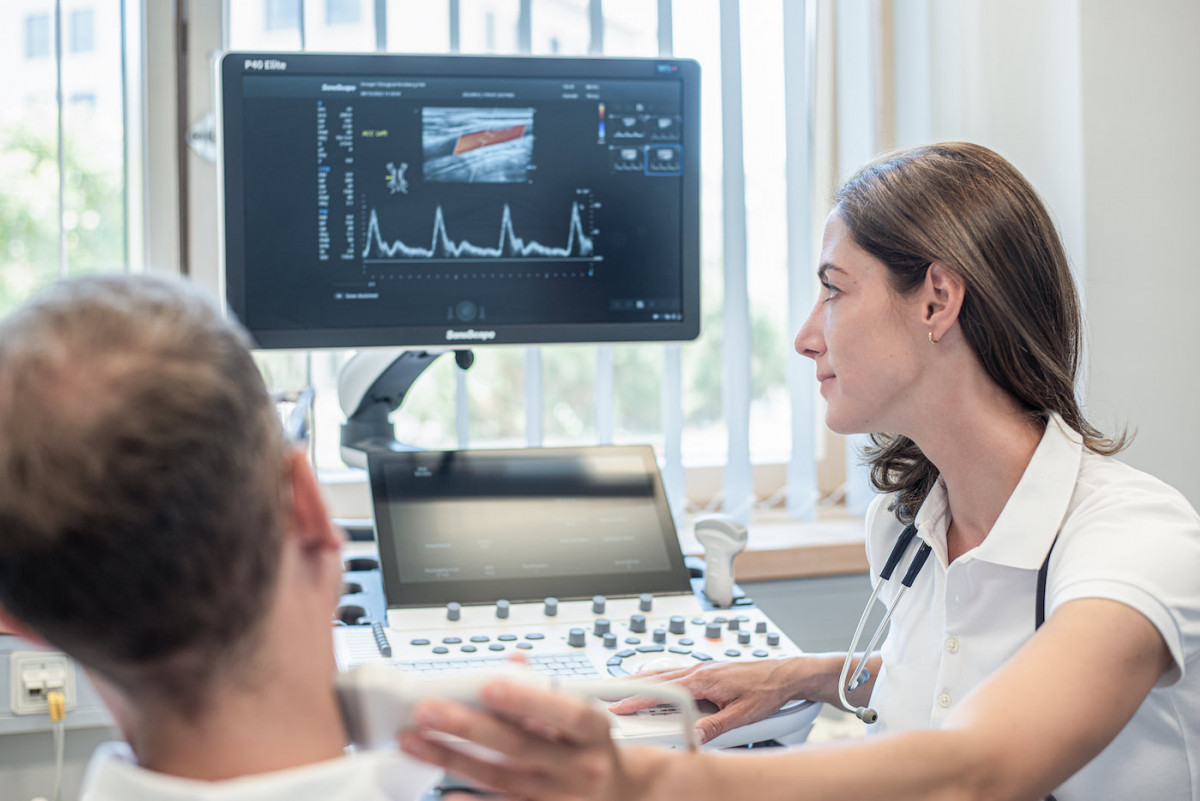
With the most modern equipment, we can provide you with an optimal and professional clarification of your complaints and carry out all preventive examinations. In addition to ultrasound examinations, the following further diagnostic procedures are available at the Hôpital Kirchberg:

  • Ankle pressure measurement and oscillography
  • Plethysmographic analysis of the small arteries of the fingers and toes
  • Imaging of the arteries and veins using computer tomography
  • Imaging of the arteries and veins using magnetic resonance imaging
  • Imaging of the arteries and veins using digital subtraction angiography (DSA)
  • Carbon dioxide angiography

    The Department of Vascular Surgery at the Hôpital Kirchberg is the only department in Luxembourg to have a carbon dioxide angiography that enables vascular imaging without the use of iodine-containing contrast media. This means that patients with functional disorders of the kidney or thyroid gland as well as a severe allergy to contrast media can also be diagnosed and optimally treated.

Our Partners

  • Hôpitaux Robert Schuman
  • National Institute Surgery Cardiac et de Cardiologie Interventionnelle
  • Collège Médical
  • Société Luxembourgeoise de Chirurgie Vasculaire
  • European Society for Vascular Surgery
  • European Union of Medical Specialists
  • German Society for Vascular Surgery and Vascular Medicine
  • German Society for Phlebology
  • Professional Association of Phlebologists

From aneurysm to access surgery - our treatment spectrum

Our Range of Services

Together we offer you the entire spectrum of modern vascular surgery. Here, we attach great importance to communicating the advantages and disadvantages as well as the procedure of different treatment methods to you in a comprehensible way.

The Department of Vascular Surgery at the Hôpital Kirchberg is the only institution in Luxembourg and the only foreign institution to have been awarded the "DGG Vascular Centre" certification by the German Society for Vascular Surgery.

The Department of Vascular Surgery at the Hôpital Kirchberg also holds the "Vein Competence Centre" certification of the German Society for Phlebology and the Professional Association of Phlebologists.

Your Patient Manual

Dear patients,
your active involvement in the treatment process is important and valuable to us!
The following patient handbook therefore provides you with important information about your medical condition and shows you what questions you can ask us and what you yourself can do to maintain or regain your health.
If any questions remain unanswered, please feel free to contact us at any time!

Information on vascular access for dialysis

Dear patient,

We would like to provide you with some information so that you can optimally prepare yourself for the planned vascular access for blood-cleansing (dialysis).

General information:

You have been diagnosed with the need for dialysis treatment, which is usually carried out three times a week (cycle A: Monday, Wednesday, Friday; cycle B: Tuesday, Thursday, Saturday). In order to be able to guarantee blood-cleansing, you need a permanent vascular access. This vascular access represents your lifeline and ensures the vital blood washing.

Types of vascular access:

The method of first choice for such vascular access is the open surgical creation of a so-called arterio-venous fistula (AV fistula) on the forearm. Here, a short-circuit connection is made between an artery (oxygen-rich blood, high pressure) and a superficial, easily accessible vein (oxygen-poor blood, low pressure). Due to the low pressure in the vein, the blood flows with little resistance from the artery into the vein, which then enlarges in diameter and thickens in the area of the vein wall in the following weeks and months. This process is called maturation (maturation). This enlarged vein (so-called shunt vein) is easily visible and palpable and can therefore be easily pricked with needles during blood washing.

The arm used is primarily the non-dominant arm, i.e. the left arm in the case of right-handers (and vice versa). Maturation takes between 8 and 12 weeks, depending on the initial diameters of the artery and vein. Therefore, the ideal time to create an AV fistula is when the patient does not yet need dialysis, but will foreseeably need it in the medium term.

If the artery and/or vein on the forearm is not suitable for the creation of an AV fistula because it is too slender or pre-diseased (vascular calcification/atherosclerosis), the short-circuit connection can also be made in the crook of the arm, on the upper arm or on the thigh.

In some cases, there is no suitable vein in the entire arm, but there is a suitable artery. In these cases, a plastic prosthesis inserted under the skin can be connected to an artery and vein and then used for blood washing. Maturation is usually not necessary. However, the surgical wounds should have healed before being used for the first time in blood-cleansing.

Both the AV fistula and the plastic prosthesis are associated with increased cardiac stress due to the greatly increased backflow of blood to the heart. In such cases, a so-called atrial catheter can be inserted. A catheter is inserted into a large vein leading to the heart through two small incisions in the neck and below the collarbone. The tip of the catheter comes to rest in the right atrium of the heart. An atrial catheter can be used directly after the operation, which is why it is also very suitable if dialysis is necessary at short notice and vascular access to the arm has not yet been planned. It can also be used to bridge the several-week maturation period of an AV fistula.

In principle, any vascular access avoiding foreign material (plastic) should be favoured as it offers considerable advantages in terms of avoiding infections as well as durability.

Consultation with your specialist:

During a detailed and confidential consultation - including an ultrasound examination of the veins - you will learn everything you need to know about the necessary preparation, the course of the operation, aftercare and possible risks and complications of the planned procedure. If you have any further questions, you are of course welcome to contact us again.

You should be able to answer the following questions:

  • Have you had dialysis access in the past? If so, when and what exactly was done?

  • What is your health status? Tell us about any illnesses, accidents or chronic conditions. Is there any heart disease? Do you have an acute infection (COVID, hepatitis, flu)?

  • Do you regularly take medication? If so, which ones and in what dosage?

  • Are you taking medication to thin your blood? If yes, which ones and in which dosage?

  • Do you suffer from allergies?

  • Do you have any blood clotting disorders?

Pre-inpatient preparation:

Before your operation, you will receive prescriptions by post for a blood test and a COVID-PCR test (may change depending on the current infection situation). You will be informed by the anaesthesiology department of the hospital about an appointment for anaesthesia information. The COVID PCR test must be carried out 24-48 hours before your admission to hospital. Please bring the negative result with you as a printout or digitally to your admission to the hospital. If the test is positive, we ask you to stay at home and inform us immediately by telephone about the positive test result. We will organise an alternative date for your operation as soon as possible.

We will also send you a surgical information sheet about the surgical procedure. We kindly ask you to read and complete this form carefully. Please be sure to bring the information sheet with you to the operation. If there are any questions, we will of course clarify them before your operation.

Important: If you are planning to have a vascular access in your arm, blood samples must not be taken from the affected arm in the weeks before the operation!

Inpatient stay:

Every type of dialysis access is performed in our hospital under inpatient conditions and in cooperation with the doctors of the Department of Nephrology. The inpatient admission usually takes place on the day before the operation, unless the patient is already in hospital due to the kidney disease.

The creation of a vascular access in the arm can be carried out under general anaesthesia or regional anaesthesia (plexus anaesthesia), depending on the existing pre-existing conditions and the patient's wishes. In the latter case, the patients are awake and only the affected arm is under anaesthesia, so that you do not feel any pain. You decide which method should be performed on you together with us and the anaesthetist.

An atrial catheter is usually inserted under general anaesthesia.

If an AV fistula is inserted, it is usually possible to discharge you the following day due to the surgical wounds. If you have had an atrial catheter implanted, its proper functioning will be checked before discharge in the course of an initial blood-cleansing.

After the insertion of a plastic prosthesis or after corrective surgery on existing dialysis lines, you should expect a stay of 5-7 days. Depending on whether the doctors of the Department of Nephrology have planned further examinations and treatments for you, the inpatient stay may be longer.

Aftercare:

You will receive prescriptions from us for plasters, painkillers and, if necessary, an outpatient nursing service to carry out the daily dressing changes.

A wound check including the pulling of the skin sutures will take place in the consultation hours of the surgeon who operated on you, usually on the 10th-12th day after the operation. You will automatically receive the appointments before discharge.

As a rule, showering is only allowed after the skin stitches have been removed!

Should you experience fever, chills, severe pain or fluid secretion in the wound area during the first few days after the operation, please call our office and come in for a check-up.

The operated arm must be rested for about two weeks.
Blood pressure measurements and blood samples must not be taken from the operated arm!

Due to the swirling blood flow in the shunt vein, a characteristic "buzzing of the shunt vein" occurs already initially after the creation of an AV fistula, which can be felt even by laypersons without prior medical knowledge.At home, the buzzing of the shunt vein should be checked several times a day by you or your relatives in order to be able to recognise possible problems at an early stage.

The maturation of the shunt vein can be accelerated by muscle work of the hand, for example by repeatedly squeezing a rubber ball. Don't worry, you can't do anything wrong here, too much exercise is not possible!

The vascular access is regularly assessed by us as well as your attending kidney specialist (nephrologist). Together we will decide when the vascular access can be used for the first time.

Despite correct puncture technique and optimal care of the vascular access, surgical corrections are often required over time due to the constant manipulation during dialysis.

Constrictions (stenoses) and occlusions (thromboses) of the shunt vein or the plastic prosthesis can occur, which have to be treated surgically or by means of catheter techniques (angioplasty).

Occasionally, dilatations of the shunt vein (aneurysms) can occur, which do not have to be corrected in every case. However, correction is necessary if the dilatation has reached an appropriate size or if there is prolonged post-operative bleeding after blood washing.

In case of very high blood flows in the shunt vein, a so-called throttling may become necessary in order to avoid a circulatory disorder of the hand and/or an overload of the heart.

Our primary goal is always to preserve an AV fistula for as long as possible, since your body's own available vein material is limited and atrial catheters and plastic prostheses must always be considered secondary.

Through our many years of work at highly specialised shunt centres in the past, we have been able to acquire extensive and excellent expertise in the surgery of dialysis accesses.Any problem with your dialysis access will be solved by us in close and valued cooperation with our colleagues in renal medicine around the clock and as quickly as possible in order to be able to guarantee your vital dialysis again!

Information about an abdominal aortic aneurysm

Dear patient,

We would like to provide you with some information about the enlargement of your abdominal aorta, the so-called abdominal aortic aneurysm, so that you can prepare yourself optimally for the examination or treatment planned with us.

General information:

We speak of an artery dilation, a so-called aneurysm, when the diameter of the vessel is more than twice the normal diameter of the vessel.

In the area of the aorta, such aneurysms occur most frequently in the abdominal cavity below the renal arteries (infrarenal aortic aneurysm). The normal diameter of the abdominal aorta is up to 2.5 cm.

The most common risk factors for the formation of an aneurysm are vascular calcification (arteriosclerosis), high blood pressure, smoking and male gender. More rarely, aneurysms develop as a result of hereditary connective tissue diseases or as a result of infections.

As with a balloon, the wall of the aneurysm becomes thinner and weaker with increasing diameter until the aneurysm finally bursts. This is almost always accompanied by life-threatening blood loss.

The vessel dilatation usually develops very slowly over several years and is usually not noticed by the patient. Up to 80% of those affected have no symptoms at all. In some cases, there is abdominal or back pain, which is always a warning sign that the aneurysm is about to burst.

Prevention:

Due to the high mortality rate of a rupture of the abdominal aorta, the primary goal is to detect an abdominal aortic aneurysm at an early stage so that appropriate treatment can be initiated.

A simple, painless, X-ray-free but nevertheless extremely safe examination method is the ultrasound examination of the abdominal cavity. Because men are more likely to develop an abdominal aortic aneurysm, professional societies recommend screening in the following population groups:

  • Men over the age of 65

  • Women over the age of 65 if they are current smokers or have smoked in the past

  • 1st degree relatives (siblings, son/daughter etc.) of patients who have already been diagnosed with an abdominal aortic aneurysm

We are happy to offer you this highly efficient examination during our consultation hours. Please feel free to contact us at any time! The good thing is that if such an aneurysm has been ruled out at an age >65 years, no further examinations are usually necessary.

In the case of preliminary stages of an aneurysm (2.5-3cm in diameter) or aneurysms smaller than 5cm, regular control examinations by means of ultrasound or a supplementary computer tomography are recommended, depending on the size.

There are no special medicines that prevent the development of an abdominal aortic aneurysm.

However, you as a patient can make a significant contribution to preventing or at least slowing down the development of an aneurysm through a healthy lifestyle, regular physical activity, weight reduction, optimal blood pressure control and, above all, by refraining from smoking.

Treatment:

Whether treatment is necessary is decided on the basis of the size and the growth rate of the aneurysm: Treatment is recommended from a diameter of 5.5cm in men or 5.0cm in women, or if the aneurysm grows at a rate of 5mm per year. In the presence of abdominal or back pain, treatment is indicated regardless of size.

The type of therapy depends primarily on the anatomy of the vessel, the patient's age and secondary diseases.

There are two main therapeutic procedures available:

In open surgical treatment, the dilated abdominal aorta is clamped above and below the aneurysm via an abdominal incision under general anaesthesia, the aneurysm is cut open and the aorta is then replaced by a tubular or Y-shaped plastic prosthesis. The shape of the prosthesis depends on whether any abnormal pelvic arteries need to be replaced in addition to the aorta. After the plastic prosthesis has been inserted, the blood flow is released and the abdomen is closed again.

This operation has been established for decades since it was first performed in 1951 and is still considered a standard procedure today.

In the second treatment method (endovascular procedure), which has been available since the beginning of the 1990s, a sheathed tubular wire mesh (a so-called stent graft prosthesis) is inserted into the abdominal aorta via both inguinal arteries under X-ray fluoroscopy and anchored in the healthy sections of the aorta located before and after the aneurysm. In this case, the pathologically changed aorta remains intact, but the aneurysm itself is excluded from the blood flow by the stent-graft prosthesis. If the prosthesis is correctly seated, the artery wall is no longer exposed to increased pressure and is no longer at risk of bursting.For this treatment, we have one of the most modern X-ray facilities in Europe at our disposal, thanks to a cooperation with the National Institute for Cardiac Surgery and Interventional Cardiology (INCCI).

The advantages of the stent-graft prosthesis over open surgery include less strain on the cardiovascular system, less pronounced pain after the operation and a shorter hospital stay.

The disadvantage of the stent graft prosthesis, however, is that certain anatomical conditions are necessary, which is why some patients are not eligible for this form of treatment. In addition, close follow-up checks by means of computer tomography and ultrasound are necessary, as the stent graft prosthesis can slip or leak in the course of time. In this case, there is again a relevant risk that the aneurysm will burst and lead to death.

In contrast, open surgery can always be performed from a surgical-technical point of view, but not all patients are suitable for this method due to certain pre-existing conditions.

We will be happy to advise you on which surgical method is suitable for your condition!

Open surgery is always performed under general anaesthesia. You should plan on staying in hospital for 7-12 days after the operation. We also recommend follow-up rehabilitation after discharge from hospital to further improve your mobility and physical fitness.

The endovascular surgical method can be performed under local or general anaesthesia, depending on age, secondary diseases, extent of the aneurysm disease and condition of the groin vessels. Most patients prefer a general anaesthetic. As a rule, you can leave the hospital about 5 days after the operation. Due to the small surgical accesses and the generally gentle surgical technique, follow-up rehabilitation is usually not necessary.

Aftercare:

The wound checks - including the pulling of the skin sutures - take place in the consultation hours of the surgeon who operated on you, as a rule on about the 5th-7th and 12th-14th day after the operation. You will automatically receive your appointments before discharge. Showering is usually only allowed after the skin stitches have been removed!

Normal physical activities and minor sports (e.g. cycling, hiking) can usually be carried out again safely 2 weeks after the operation.

You should refrain from more strenuous physical activities (heavy physical work, sports, swimming, sauna, etc.) for 8 weeks after an open operation and for 4 weeks after an endovascular operation.

After an open operation, follow-up examinations by ultrasound are usually sufficient. In case of complaints or the need for a more precise diagnosis, a computer tomography with contrast medium may also be necessary.

After endovascular treatment, structured follow-up examinations are necessary every 6 to 12 months in order to be able to detect any leakage of the stent graft prosthesis at an early stage. In the event of a leak, an individual decision is made as to whether further surgery is necessary, depending on the type and extent of the leak.

As a rule, you will need a mild blood thinner, a so-called platelet aggregation inhibitor (e.g. Cardioaspirin/Aspirin/ASS or Clopidogrel/Iscover/Plavix), for a long time after the operation.If you have already been taking blood thinners before the operation, for example due to heart disease, you can usually simply continue to take this medication.Equally important are optimal blood pressure control with medication and regular monitoring of blood lipid levels. If necessary, you will be prescribed a lipid-lowering agent.

Information on chronic wounds

Dear patient,

We would like to provide you with some information so that you can optimally prepare yourself for the therapy of a chronic wound you are about to receive.

General information:

Chronic wounds are of considerable socio-economic importance and usually pose great challenges to patients and their doctors. The restriction of the quality of life and the suffering of the patients is usually high and the care requires a high nursing and medical effort.

A wound is considered "chronic" if it does not show any healing tendency within 6-8 weeks despite causal and appropriate local treatment.

The most frequent causes for the development and maintenance of chronic wounds are the late effects of deep vein thrombosis, a functional impairment (insufficiency) of the venous valves, a circulatory disorder of the legs (atherosclerosis), diabetes mellitus or chronic pressure.

Chronic wounds most frequently occur on the lower leg and foot and are referred to here as "open leg", leg ulcer or foot ulcer. If a chronic wound is due to diabetes, it is usually called "malum perforans".

Efficient wound therapy always begins with clarifying the cause of the disease. This is almost always done by means of painless diagnostics (blood tests, blood pressure measurement and ultrasound examination of the legs).

Therapy depends largely on the underlying cause and ranges from consistent compression therapy and optimisation of blood sugar to restoration of arterial circulation, surgical wound cleansing and vacuum sealing therapy to hyperbaric oxygen therapy and modern, highly specific wound dressings.

Restoration of blood circulation:

The most dangerous are chronic wounds due to arterial circulatory disorders. These are statistically associated with a high amputation rate. Preservation of the limb and thus mobility is the highest goal of successful wound therapy!

In order to diagnose an arterial circulatory disorder, it is first necessary to measure the blood pressure in the arms and legs and to perform an ultrasound examination. In most cases, depending on the kidney function, a computer tomography or magnetic resonance tomography is also carried out to show the arteries that supply the leg with oxygen-rich blood.

If narrowing or occlusion of the arteries is found, an individual therapy strategy tailored to your needs will be worked out. This may include open surgical treatment (atherectomy), vascular dilatation by means of catheters (angioplasty) and severing of the sympathetic nerve (sympathicolysis).

Compression therapy:

If a venous disease, such as the late stage of a leg vein thrombosis or an inability of the deep venous valves to function, is the cause of the chronic wound, consistent compression therapy of the legs with compression stockings of compression class II is a decisive component of the treatment success, without which healing of the wound cannot be expected. Alternatively, elastic bandages can be used.

The pressure exerted by the stockings and bandages (ankle pressure of 23-32 mmHg) relieves the veins and improves the transport of blood from the leg to the heart.

Surgical wound cleansing (debridement):

In surgical wound cleansing, dead or inflamed tissue is removed under locally sterile conditions using tweezers, a sharp-edged spoon or a scalpel. Depending on the patient's pain tolerance and the extent of the wound, this can be done without anaesthesia, under local anaesthesia or under general anaesthesia in the operating theatre. The wound can also be cleaned with a jet of water applied under high pressure (so-called jet lavage).

Regardless of the procedure, repeated surgical debridement may be necessary.

In the case of underlying arterial or venous disease, surgical wound cleansing should always be considered as a complement to improved circulation or consistent compression therapy.

Vacuum sealing therapy:

Vacuum sealing therapy has proven to be very effective in healing chronic wounds. After surgical wound cleansing, the wound is covered airtight with a special dressing consisting of a large-pored sponge and a foil. Wound fluid is drained continuously or at intervals into an associated pump via a thin tube, creating a negative pressure in the wound area which improves the blood supply to the wound and stimulates the damaged tissue to grow.

The negative pressure on the wound can be regulated. The dressing is usually changed every 3 to 5 days, the wound inspected, surgically cleaned again if necessary and the sponge and tubing system replaced.

However, the connected pump slightly restricts mobility and makes noise from time to time during suction, which some patients find annoying.

Hyperbaric oxygen therapy:

In this treatment method, 100% pure oxygen is administered to the limb affected by the chronic wound using a special chamber under positive pressure.

The combination of 100% oxygen and the simultaneous application of hyperbaric pressure allows for very high "oxygen dosages" (oxygen partial pressures) in the blood and tissues, which has a number of positive effects on wound healing, such as reducing water retention in the tissues, promoting new blood vessel formation (angiogenesis) and displacing harmful metabolic end products and toxins.

There are indications that hyperbaric oxygen therapy can improve wound healing, especially in the case of damage to the smallest blood vessels (microangiopathy), as occurs in diabetes, chronic kidney disease or also in atherosclerosis.

Split thickness skin grafting:

If a wound is so large that it cannot close by itself, a split-thickness skin graft (split-thickness skin transplantation) is considered.In this procedure, the upper parts of the skin are removed from a healthy part of the body, usually the thigh, using a special device called a dermatome and transplanted onto the clean and pre-treated wound.As a rule, the thickness of the skin flap is 0.08-0.2inch.

The removed skin leaves only a superficial "graze wound" at the removal site, which heals within 2 to 3 weeks with the application of special wound dressings and ointments.

In order to keep the area of skin removed as small as possible, diamond-shaped incisions are made in the removed skin with the help of a knife roller before transplantation. This is why the treatment procedure is strictly speaking called mesh grafting.

This allows areas 1.5-3x the size of the piece of skin removed to be covered. In addition, a mesh graft has the advantage that wound fluid can drain through the openings in the skin. Mesh split skin usually heals very well by "filling in" the gaps in the skin with the patient's own skin.

The disadvantage of this procedure is that the meshes can be recognised as a typical pattern even after years.

Wound dressings:

Wounds should be covered with special dressings to protect them from irritation, dirt and germs.

Classic dressing materials, such as gauze and gauze compresses and non-woven fabrics are called inactive wound dressings. They can be used to apply disinfectant solutions and to cleanse the wound with saline solution. As wound dressings, they absorb blood and wound secretions, but they have the disadvantage that they tend to stick together when more fluid is secreted and can then only be removed from the wound with difficulty and possibly painfully.

Since wound healing is usually facilitated in a moist environment, there are various so-called interactive wound dressings such as hydrogels, hydrocolloids, silver or alginate-containing wound dressings, foam dressings, foil and gauze to prevent the wound from drying out.Interactive wound dressings usually remain on the wound for a few days and should be changed when they have become noticeably soaked with wound secretions or have slipped.

Antibiotics:

If a wound looks infected or bacteria can be detected in a swab of the wound, antibiotics should be administered in addition to wound care. Whether these are administered in tablet form or into the vein depends on the severity of the infection and the type of bacteria. Antibiotics that are easily absorbed by tissue and bone are usually used.

In contrast, evidence of normal bacterial skin flora in an externally uninfected-looking wound is not a reason for antibiotic therapy. Antibiotics should always be administered with restraint to protect against bacterial resistance and multidrug-resistant germs.

Pain management:

Chronic wounds often cause chronic pain. We therefore work together with the "pain service" of the Hôpital Kirchberg, consisting of specialised doctors and nurses to treat your pain early and optimally.

Information about surgery on varicose veins

Dear patient,

We would like to provide you with some information so that you can prepare yourself optimally for the varicose vein operation planned for you.

Consultation with your specialist:

During a detailed and confidential consultation, including a painless ultrasound examination of the veins, you will learn everything you need to know about the necessary preparation, the course of the operation, aftercare and possible risks and complications of the planned procedure. Should further questions arise, you are of course welcome to contact us again.

After we have answered all your questions comprehensively, we also need some important information from you in order to be able to plan the upcoming operation optimally.

You should be able to answer the following questions:

  • Have you already had surgery on varicose veins in the past? If so, when and what exactly was performed?

  • What is your health status? Tell us about any illnesses, accidents or chronic conditions. Is there any heart disease? Do you have an acute infection (COVID, hepatitis, flu)?

  • Do you regularly take medication? If so, which ones and in what dosage?

  • Are you taking medication to thin your blood? If yes, which ones and in which dosage?

  • Do you suffer from allergies?

  • Do you have any blood clotting disorders?

Pre-operative preparation:

Before your operation you will receive a prescription by post for thigh compression stockings of compression class II, which you can have made to measure in a specialist medical shop. These compression stockings do not have to be worn before the operation. However, we do ask you to bring the compression stockings with you to the hospital for your operation.

You will also receive prescriptions from us for a blood test and a COVID-PCR test (may change depending on the current infection situation). You will be informed by the anaesthetics department of the hospital about an appointment for anaesthetic information. Please bring your blood results with you to this appointment.

The COVID PCR test must be carried out 24-48 hours before your admission to hospital. Please bring the negative result with you as a printout or digitally to your admission to the hospital. If the test is positive, we ask you to stay at home and inform us immediately by telephone about the positive test result. We will organise an alternative date for your operation as soon as possible.

We will also send you a surgical information sheet about the surgical procedure. We kindly ask you to read and complete this form carefully. Please be sure to bring the information sheet with you to the operation. If there are any questions, we will of course clarify them before your operation.

The day of the operation and different operation methods:

The operation is usually done on an outpatient basis. This means that you come to the hospital fasting on the morning of the operation and are allowed to leave the hospital on the same day, about four hours after the operation. Please bear in mind that you are not allowed to drive on your own on the day of the operation.

In the case of certain pre-existing conditions, special medication (especially blood thinning medication), certain varicose vein operations in the past history or at your request, we will perform the operation under in-patient admission to the hospital. You will be admitted to hospital in the afternoon of the day before the operation. As a rule, you will be discharged on the first day after the operation.

In order to ensure an optimal aesthetic result, the varicose veins are marked in a standing position before the operation. The operation itself takes place under general anaesthesia, so you will not feel any pain during the procedure.

In the so-called Babock vein stripping procedure, the non-functioning sections of the saphenous vein, which extends from the foot to the groin or to the knee, are removed.The operation itself begins with an incision in the groin or in the back of the knee. All superficial veins that join the deeper veins are tied off. Then, through a tiny incision in the ankle, lower thigh or thigh (above the so-called lower insufficiency point, i.e. the lowest point of dysfunction of the venous valves), a probe is inserted into the diseased section of the vein and the dysfunctional sections of the saphenous vein are then pulled.

Side branch varicose veins are removed via a so-called miniphlebelomy using tiny punctures of the skin.

In radiofrequency ablation, the patient is first given a so-called tumescent anaesthetic (a mixture of saline solution and local anaesthetic) in the subcutaneous fatty tissue for heat protection and local anaesthesia. The saphenous vein in the ankle or lower leg is then punctured with a fine needle to insert the catheter. This catheter emits radiofrequency energy from its tip into the vein wall, heating it up to 120 degrees Celsius and causing the vessel to contract and shrink. During the operation, the position of the catheter is checked by ultrasound, so that an advance of the catheter into the deep vein system and thus a heat damage of the same is consistently avoided.

The wounds in the groin and back of the knee as well as on the ankle are usually sutured, smaller stitches after removal of side branch varicose veins are only glued with suture strips.

After the operation, the operated leg is wrapped tightly and elastically under anaesthesia. Drains to drain off wound fluid are usually only inserted in complex operations or multiple previous operations on the varicose veins.

After the operation you will be monitored by the anaesthetist for another 4 hours in the recovery room and then discharged or transferred to the ward.

You will be able to eat and drink again shortly after the operation. On the day of the operation you should already get up and - as far as it is tolerated due to the pain - also move around.

Before you are discharged, your surgeon will check the surgical wounds and answer any questions you may have about the operation. In addition, the elastic wrapping will be removed and the custom-made class II compression stockings you brought with you will be put on. In the case of an operation with an overnight stay in hospital, a dressing change with wound control and, if necessary, the pulling of a drainage is carried out on the first day after the operation.

Aftercare:

You will receive prescriptions from us for plasters, painkillers and injections to prevent thrombosis (heparin injections). The heparin injections must be administered for another 10 days after the operation. The application is simple and is usually learned quickly. The nursing staff will support and guide you. Please do not hesitate to contact us if you do not feel confident to administer the syringes yourself. In this case, we will be happy to write you a prescription for an outpatient nursing service.

The bandages in the groin and the back of the knee should be changed daily. The bandages over the smaller stitches should only be changed if they are more saturated with blood or wound secretions. The suture strips should be left in place if they do not fall off by themselves.

Painkillers should be taken according to the symptoms. Compression stockings should be worn consistently during the day and night for 7 days, then only during the day for another 5 weeks. Compression therapy is an enormously important part of the success of the therapy and should not be underestimated! As a rule, showering is only allowed after the skin threads have been pulled!

The wound checks, including the removal of the sutures, are carried out in the consultation hours of the surgeon who operated on you, usually on the 5-7th and 12-14th day after the operation. You will automatically receive your appointments before discharge.

Should you experience fever, chills, severe pain or fluid secretion in the wound area in the first few days after the operation, please come to our clinic for a check-up after a brief telephone consultation via our secretary's office.

Normal physical activities and minor sports (e.g. cycling, hiking) may be resumed as far as possible immediately after the operation. Everything that is not perceived as painful is permitted. You should refrain from extreme physical stress (heavy physical work, sports, swimming, sauna, etc.) for 2 to 4 weeks.

Everyday activities and occupations in a primarily seating position can usually be performed again one week after surgery. In the case of standing or physically strenuous occupations, the downtime is up to 14 days.

Direct sunlight and solarium should be avoided for 6 weeks, as otherwise cosmetically unpleasant hyperpigmentation (brownish discolouration) may occur in the area of the scars.

Depending on individual tolerance, the fitted compression stockings can continue to be worn after the 6 weeks are over. In principle, there is no such thing as too much compression therapy and the more severe the varicose vein condition was before the operation, the more sensible it is to wear the compression stockings for a long time. We also recommend wearing compression stockings during long car journeys or air travel to reduce water retention in the legs and to prevent thrombosis.

Regular follow-up checks using ultrasound are not usually necessary after varicose vein surgery. However, since the predisposition to develop varicose veins persists, it is not uncommon for varicose veins to reappear, albeit usually less prominent. Should cosmetically disturbing or promblematic varicose veins (with thrombosis, inflammation, bleeding) reappear in you months or years later, another varicose vein operation is not a problem.Therefore, please feel free to contact us!

Information on arterial vascular disease of the legs

Dear patient,

We would like to provide you with some information on your present arterial vascular disease of the legs, so that you can prepare yourself optimally for the examination or treatment planned with us.

General information:

Arterial vascular disease of the legs, also called peripheral arterial occlusive disease or "window shopper's disease" is the most common vascular disease and therefore of considerable importance. Blood can no longer flow freely due to narrowing and occlusion of the vessels, which results in an undersupply of oxygen and important nutrients to the leg.

Risk factors for this disease are smoking, high blood pressure, diabetes, elevated blood lipid levels, obesity, lack of exercise and an unhealthy diet.

Symptoms:

The disease can initially run without symptoms for a long time, even years, which corresponds to the first stage of the disease. Typical symptoms of the second stage of the disease are sore muscles in the calf or thigh muscles. Affected persons typically stop walking when the pain occurs and wait until the symptoms have subsided. In order to distract the public from the actual cause of the standstill, those affected look at shop windows, which is why it is commonly referred to as " window shopper's disease". The second stage is further subdivided into stage IIa with a pain-free walking distance of over 200 metres and stage IIb with a pain-free walking distance of less than 200 metres.

The walking distance that can be covered without pain inevitably decreases as the disease progresses. Finally, the pain occurs even without movement, i.e. at rest, especially at night. Affected persons are then in the third stage of the disease and initially alleviate the pain unconsciously by letting the leg hang down from the bed, which slightly improves the blood supply in the foot. Nevertheless, the pain occurring at rest is an absolute warning signal for a critical reduced blood supply in the leg and should lead to an immediate presentation to a specialist in vascular surgery.

It becomes particularly problematic when a nerve disorder (polyneuropathy), which is most common in diabetes, is present at the same time and the affected person does not feel the pain in the legs and feet. The disease then progresses unnoticed to the fourth and final stage, when tissue destruction occurs. This results in black and open spots on the toes or feet. If the affected person gets a wound, for example during foot care or due to a minor injury, this wound will not heal.

In the case of a sudden deterioration or after previous treatments, acute, severe pain in the leg can also occur, often in connection with a feeling of cold and numbness as well as paleness. In this case, a doctor should be consulted immediately.

Therapy:

Therapy options include open surgical treatment (atherectomy), vessel widening by means of balloon catheters (balloon angioplasty), if necessary with insertion of a vessel support (stent angioplasty), severing of the sympathetic nerve (sympathicolysis) and alleviation of the symptoms by painkilling and circulation-enhancing drugs.

The preservation of the limb and thus of mobility is the highest goal of a successful therapy! The earlier the disease is recognised, the higher the probability of improving and preserving the ability to walk in the long term.

If a longer walking distance of, for example, several hundred metres is still maintained, the treatment method can initially be purely conservative with medication and so-called structured gait training. If the increase in walking distance is still insufficient for those affected, invasive therapy can be considered.

The basic principle is always: do not operate too early and too extensively, because the disease progresses steadily even with optimal drug therapy and the surgical options on one leg are limited in principle.

If the disease is only diagnosed at a late stage and there are already open areas on the foot, the probability of amputation in the following years is unfortunately very high.

An important part of the therapy is also the consistent control and improvement of the risk factors, i.e. a good blood pressure and blood sugar control, a reduction of elevated blood lipid values, a weight reduction and an increase in exercise and, most importantly, a strict ban on smoking!

Also, window shopper's disease must be seen as a "marker disease" for other conditions, such as coronary artery disease and carotid artery calcification. Patients with advanced arterial occlusive disease of the legs have a high risk of suffering a heart attack or stroke in the following years. We urge you to always be aware of this fact and to take your condition seriously!

By asking simple questions and using diagnostic tools such as an ultrasound examination of the neck vessels, these diseases can be detected and treated at the same time! We work closely with angiologists, diabetologists, cardiologists and neurologists.

Together with you, we do everything we can to preserve your leg and your mobility, your independence and your self-determination for as long as possible!

Training

Due to the extensive treatment spectrum, we at the Hôpitaux Robert Schuman have full authorisation for further training to become a specialist in vascular surgery, granted by the Medical Association of Saarland (Germany).

The Hôpitaux Robert Schuman is also an academic teaching hospital of the Mannheim Medical Faculty of the University of Heidelberg (Germany) and the Medical University of Innsbruck (Austria). In this context, German and Austrian medical students can complete part of their final year of study ("practical year") in the Department of Vascular Surgery at the Hôpital Kirchberg.

Likewise, part of the specific training in general medicine (Formation Spécifique en Médecine Générale, FSMG) at the University of Luxemburg can be completed in the Department of Vascular Surgery at the Hôpital Kirchberg.

  • Medical Association of Saarland
  • Mannheim Medical Faculty of the University of Heidelberg
  • Medical University of Innsbruck
  • University of Luxemburg